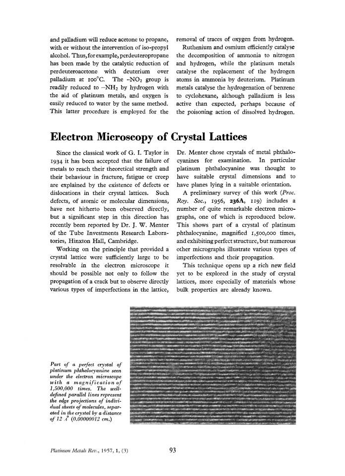
Electron Microscopy of Crystal Lattices
Journal Archive
Electron Microscopy of Crystal Lattices
Since the classical work of G. I. Taylor in 1934 it has been accepted that the failure of metals to reach their theoretical strength and their behaviour in fracture, fatigue or creep are explained by the existence of defects or dislocations in their crystal lattices. Such defects, of atomic or molecular dimensions, have not hitherto been observed directly, but a significant step in this direction has recently been reported by Dr. J. W. Menter of the Tube Investments Research Laboratories, Hinxton Hall, Cambridge.
Working on the principle that provided a crystal lattice were sufficiently large to be resolvable in the electron microscope it should be possible not only to follow the propagation of a crack but to observe directly various types of imperfections in the lattice, Dr. Menter chose crystals of metal phthalocyanines for examination. In particular platinum phthalocyanine was thought to have suitable crystal dimensions and to have planes lying in a suitable orientation.
A preliminary survey of this work (Proc. Roy. Soc., 1956, 236A, 119) includes a number of quite remarkable electron micrographs, one of which is reproduced below. This shows part of a crystal of platinum phthalocyanine, magnified 1,500,000 times, and exhibiting perfect structure, but numerous other micrographs illustrate various types of imperfections and their propagation.
This technique opens up a rich new field yet to be explored in the study of crystal lattices, more especially of materials whose bulk properties are already known.